Figure 4.

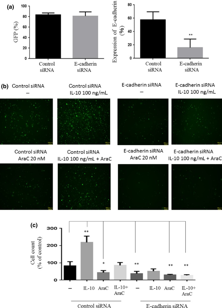
Function of E‐cadherin in leukemia cells. (a) GFP‐positive THP‐1 cells were transfected with control or E‐cadherin‐specific siRNA and analyzed for GFP and E‐cadherin expression by flow cytometry. **P < 0.01. (b) GFP‐positive THP‐1 cells were cocultured with UE6E7T‐3 human bone marrow‐derived mesenchymal stem cells and then treated with cytarabine (AraC) and/or interleukin‐10 (IL‐10). After 48 h, floating cells were removed and adherent cells were washed twice. GFP‐positive cells were analyzed by fluorescence microscopy. (c) Cell counts were undertaken using BZ‐II Dynamic Cell Count software.
