Figure 3.

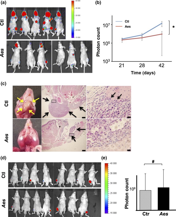
Aes suppresses metastatic spread of human PCa cell grafts to the bone in mice. (a) In vivo bioluminescence images of mice injected with luciferase‐expressing PC3 cells into the left cardiac ventricle. (b) Quantification of the bone metastatic lesions (photon counts) in mice injected with luciferase‐expressing PC3 cells as shown in (a). Aes, Aes‐expressing PC3 cells; Ctl, no‐Aes control cells. (c) Microphotographs of bone metastatic lesions in mice injected with luciferase‐expressing PC3 cells into the left ventricle. Arrows indicate metastatic lesions. Framed regions (dotted rectangles) are enlarged on the right. Scale bar; middle 500 μm, right 20 μm. (d) In vivo bioluminescence images of mice injected with luciferase‐expressing PC3 cells into femurs directly. (e) Quantification of the bone tumor lesions (photon counts) in mice injected with luciferase‐expressing PC3 cells as shown in (d). Asterisk (*) in (b) indicates statistically significant difference (P < 0.05), whereas pound (#) in (e) indicates no statistical significance.
