Fig. 5.
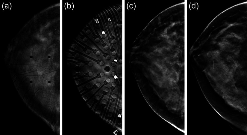
Impact of optical plates on the DBT images. (a) Top slice of breast DBT volume taken with the optical probe attached. The source fibers and mounting holes are clearly visible. (b) Bottom slice of breast DBT volume taken with the optical probes attached. The detector fibers and prisms are clearly visible. The high absorption patches are the result of small pieces of electrical tape used in the construction of the probe. They were removed subsequently. (c) Middle slice of breast DBT image taken with the optical probe attached. Faint shadows of the detector fibers can be seen. (d) For comparison, middle slice of the separately acquired clinical breast DBT image taken on the same patient (no optical components present).

