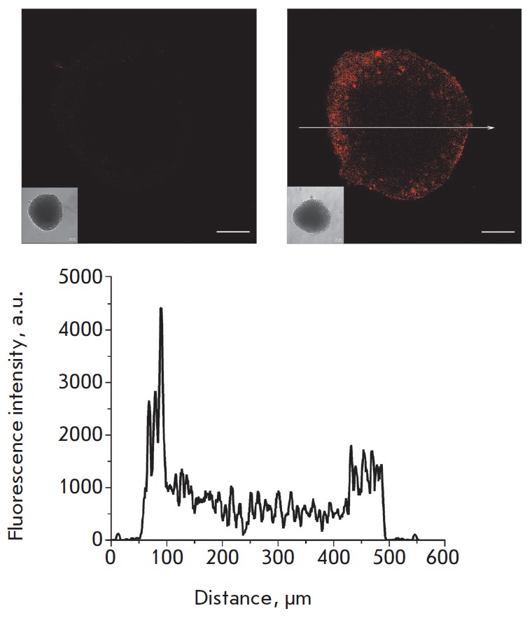
Fig. 6.

Confocal images of an unstained SKBR-3 spheroid (left) and SKBR-3 spheroid stained for 2 h with 4D5scFv- PE40 conjugated with a DyLight650 fluorescent dye (right). Bar, 100 μm. The insets show wide-field microscopy images of the same spheroids. Bottom: the fluorescence signal profile along the arrow shown in the right-hand side image.
