
Figure 1.
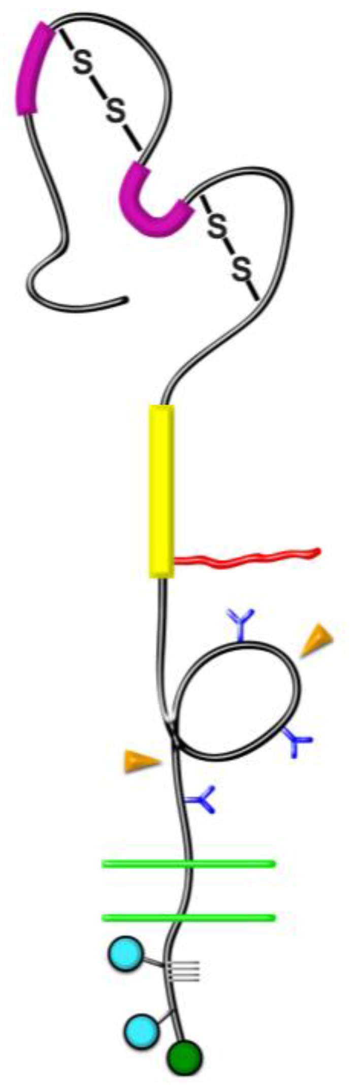
Domain structure of NG2. NG2 is a type-1 transmembrane protein with several distinct structural domains. Domain 1. Bold magenta bars = laminin G domains; S-S = disulfide bonds. Domain 2. Bold yellow bar = collagen binding domain; Irregular red line = chondroitin sulfate chain attached at S-999. Domain 3. Blue Y shapes = N-linked oligosaccharides; Orange arrowheads = sites of proteolytic cleavage. Transmembrane domain. Double green lines = plasma membrane. Cytoplasmic domain. Blue circles = sites of threonine phosphorylation at T-2256 and T-2314; Green circle = C-terminal PDZ-binding motif; Gray grid lines = proline-rich segment. Figure taken from [17].