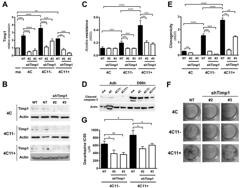
Figure 1.
Downregulation of Timp1 decreases cell survival in melanoma cells. (A,B) Timp1 mRNA expression was assessed by RT-qPCR and Western blotting, respectively, in different cell lines representing different stages of melanoma progression after transduction with viral particles containing two different shRNA sequences for Timp1 (shTimp1#2 and shTimp1#3) or control non-target shRNA (non-target, NT); (C,D) cell lines were maintained in suspension for 96 h and the number of viable cells was estimated, respectively, using MTT and analysis of cleaved caspase-3 by Western blotting; (E,F) 200 cells were incubated at 37 °C on 60 mm2 plates for 10 days. After this period, the clone formation was, respectively, quantified and visualized after toluidine blue staining; and (G) the non-metastatic 4C11− and metastatic melanoma cell line 4C11+ were treated for 48 h with dacarbazine (IC50). After this period, cell viability was analyzed by MTT assay. ma: melan-a melanocytes; 4C: pre-malignant melanocytes; 4C11−: non-metastatic melanoma cell line; 4C11+: metastatic melanoma cell line; NT: control non-target shRNA; shTimp1#2: clone 2 silenced for Timp1; shTimp1#3: clone 3 silenced for Timp1. * p < 0.05, ** p < 0.01, *** p < 0.001, **** p < 0.0001.

