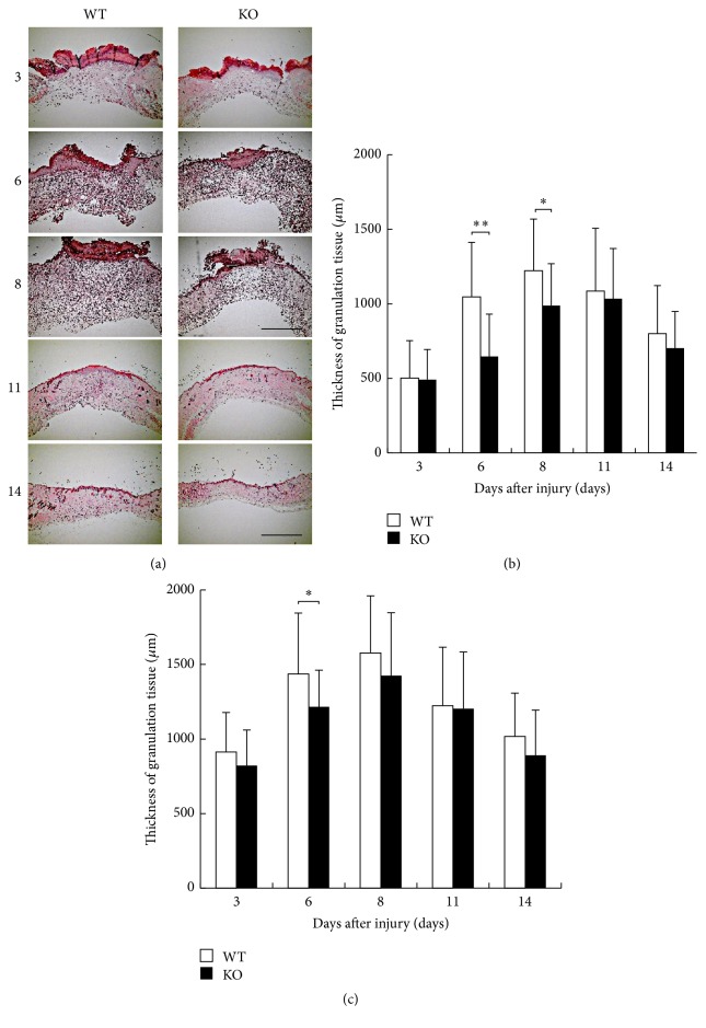
Figure 2.
(a) Histology of healing wound tissue by hematoxylin and eosin stain at the indicated time intervals in dorsal skin of inducible nitric oxide synthase (iNOS-) knockout (KO) mice and WT mice. (b) The thickness of the granulation tissue at the center zone was thinner in KO mice than in WT mice at days 6 and 8 with a statistically significant difference. (c) At the margin zone, the thickness of the granulation tissue was significantly thinner in KO mice than in WT mice at day 6. Open bars: WT, filled bars: KO (days 3, 11, and 14: n = 6; days 6, 8: n = 7 animals in each group). Mean ± standard deviation. ∗p < 0.05; ∗∗p < 0.01; bar, 1 mm.

