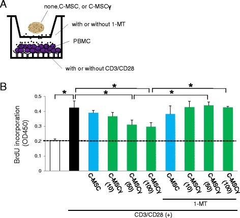
Fig. 2.

C-MSCγ attenuates T cell proliferation via its IDO activity. a Schematic figure of the co-culture system. Transwell culture plates with 0.4 μm pores were used. PBMCs were cultured in the bottom compartment of plates that were pre-coated with or without anti-human CD3 (5 μg/mL) and CD28 antibodies (2 μg/mL) to stimulate T lymphocyte proliferation. A C-MSC or C-MSCγ (10, 50, 100) was set in the upper chamber in the presence or absence of an IDO inhibitor, 1-MT (500 μM). They were co-cultured for 72 h in RMPI medium. b BrdU was added to the medium 3 h before the end of the incubation. Then, incorporated BrdU was quantified by measuring the optical density at a wavelength of 450 nm (OD450) using an ELISA kit system. Values represent means ± S.D. of four cultures. * p < 0.05: values differ significantly (t test). Similar results were obtained from three experiments. 1-MT 1-methyltryptophan, BrdU bromodeoxyuridin, CD cluster of differentiation, C-MSC clumps of a mesenchymal cell/extracellular matrix complex (C-MSC) cultured in growth medium for 3 days. C-MSCγ (10, 50, or 100) C-MSC treated with 10, 50, or 100 ng/mL IFN-γ for 24 h before the end of the culture period, PBMCs peripheral blood mononuclear cells
