
Fig. 9.
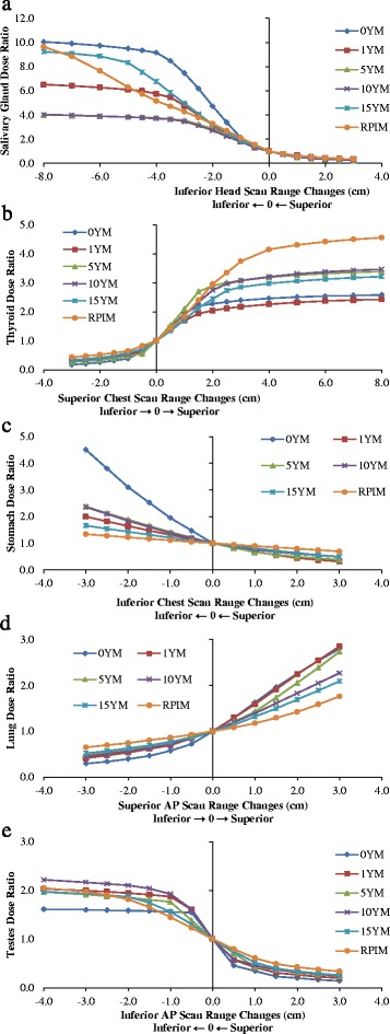
The sensitivity of doses of partially scanned organs in respect to scan range change: Head scans: a) Salivary Gland; Chest scans: b) Thyroid and c) Stomach; AP scans: d) Lungs and e) Testes; the arrows in horizontal axis title indicate the scan range change in the corresponding direction leads to increase in dose