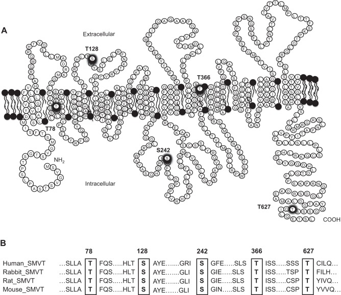
Fig. 4.
Schematic diagram showing the putative CK2 phosphorylation site in the hSMVT protein. A: the hSMVT protein is predicted [by NetPhos 3.1 Server (9)] to have five putative CK2 phosphorylation sites (thick circles) that are conserved in different species. B: conserved amino acid residues (in rectangles) in different species were identified by multiple sequence alignment using “PRALINE” software (www.ibi.vu.nl/programs/pralinewww/).

