Fig. 6.
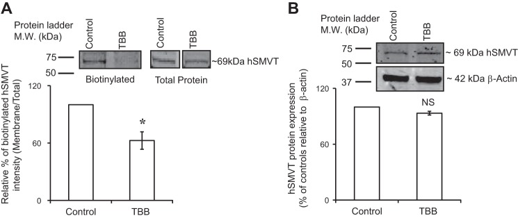
Effect of the CK2 inhibitor TBB on expression of the hSMVT protein at the surface of NCM460 cells. Cells (maintained in serum-free medium) were treated with the TBB (30 µM; 1 h), followed by biotinylation assay (see Methods). A: surface expression of hSMVT was quantified using anti-hSMVT polyclonal antibodies and was normalized relative to total cellular hSMVT level. B: total cellular hSMVT was quantified and then normalized relative to β-actin. Inset: representative images from biotinylation assay. In both Western blot analyses, identical amounts of protein (60 μg) were loaded onto gels. Data are means ± SE of at least 3 independent experiments (*P < 0.01).

