Fig. 2.
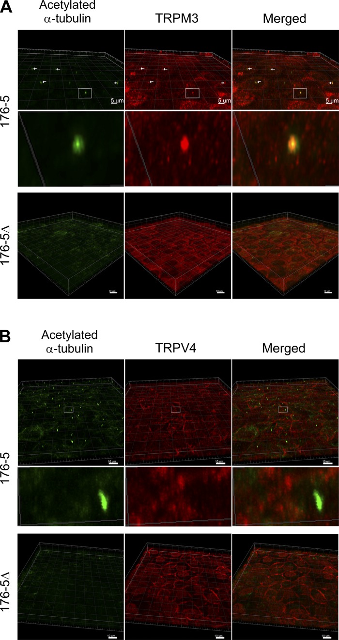
TRPM3 and TRP vanilloid-4 (TRPV4) cellular localization in 176-5Δ and 176-5 cells. A: immunofluorescence was performed using primary antibodies directed against acetylated α-tubulin (green) and TRPM3 (red), and images were overlaid (Merged). Images were obtained using a Zeiss LSM710 confocal microscope and analyzed using Imaris Scientific 3D/4D Image Analysis software. Top: in 176-5 cells, arrows indicate the presence of cilia (left), ciliary TRPM3 (middle), and colocalization (right). Scale bars = 5 µm. Middle: magnified view of the area within the white box in top row. Bottom: 176-5Δ cells were devoid of primary cilia (green, left), and TRPM3 expression was observed diffusely throughout the cells (middle and right). Scale bars = 10 µm. B: immunofluorescence was performed using primary antibodies directed against acetylated α-tubulin (green) and TRPV4 (red), and images were overlaid (Merged). Images were obtained and analyzed as described in A. Top: primary cilia were observed in 176-5 cells (green, left); cell body (possibly plasma membrane) expression of TRPV4 expression was apparent (red, middle), but no ciliary expression of TRPV4 was observed (right). Middle: magnified view of the area within the white box in top row. Bottom: 176-5Δ cells were devoid of primary cilia (green, left); cell body expression of TRPV4 resembled that of 176-5 cells (middle and right). Scale bars = 10 µm.

