Fig. 4.
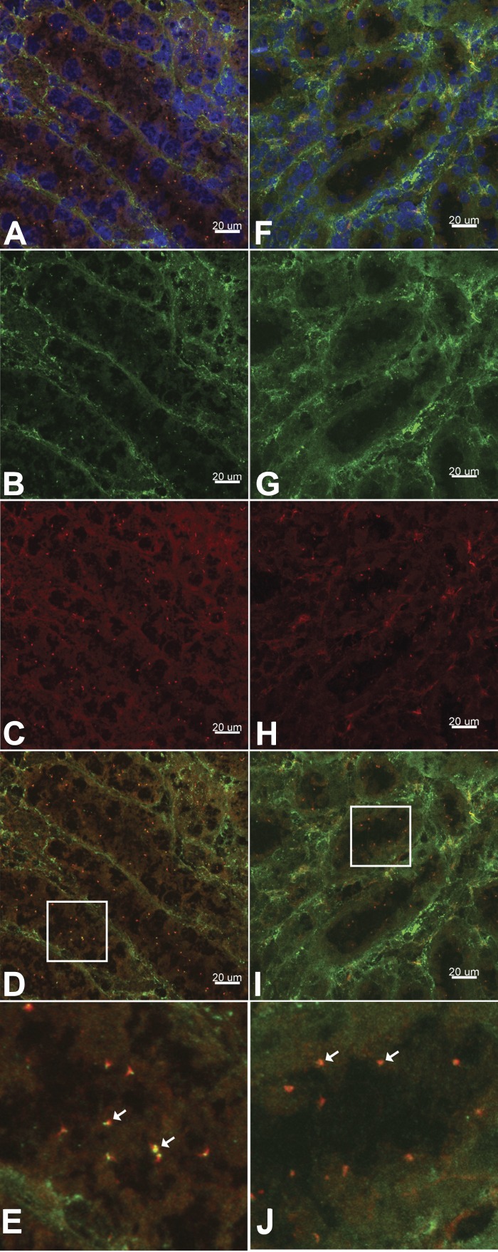
TRPM3 cellular localization in wild-type mouse kidney tissue. A–E: frozen tissue sections were subjected to immunofluorescence using primary antibodies directed against acetylated α-tubulin (green, A, B, D, and E) and TRPM3 (red, A, C, D, and E). Nuclei were labeled with DAPI (blue, A). TRPM3 coexpression with acetylated α-tubulin (yellow, D and E) indicates localization on primary cilia. F–J: frozen tissue sections were subjected to immunofluorescence using primary antibodies directed against γ-tubulin (green, F, G, I, and J) and TRPM3 (red, F, H, I, and J). Nuclei were labeled with DAPI (blue, F). TRPM3 coexpression with γ-tubulin (yellow, I and J) supports ciliary localization of the channel. Images were obtained using a Nikon A1 inverted confocal microscope and analyzed using Imaris Scientific 3D/4D Image Analysis software. E and J: magnified views of areas in white boxes in D and I, respectively. White arrows indicate coexpression.

