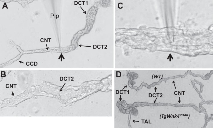
Fig. 1.
Distal convoluted tubule (DCT) preparation for the patch-clamp recording. A: tubule image showing the location of DCT1, DCT2, connecting tubule (CNT), and cortical collecting duct (CCD). B: tubule image showing the split-open DCT2 and CNT. C: image showing a patch-clamp pipette (Pip) was placed in the apical membrane of a split open DCT2/CNT. The pipette position pointed by an arrow is also indicated in A. D: images of isolated DCT/CNT from wild-type (WT) and TgWnk4PhAII mouse. Note that DCT/CNT tubule from TgWnk4PHAII mouse is hypertrophic in comparison to WT.

