Fig. 4.
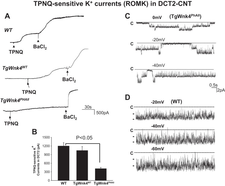
ROMK activity was inhibited in TgWnk4PHAII mice. A: set of TPNQ-sensitive K+ current traces measured with the perforated whole cell recording in WT, TgWnk4WT, and TgWnk4PHAII mice. The pipette solution was composed of 140 mM KCl, 2 mM MgCl2, 1mM EGTA, and 5 mM HEPES (pH 7.4). The membrane potential was clamped at −40 mV for the measurement of K+ currents. Addition and washout of TPNQ (400 nM) are indicated by arrows. B: bar graph summarizes the results measured at −40 mV. C: single channel recording demonstrates diminished ROMK channel activity in the apical membrane of DCT2 of TgWnk4PHAII mice. D: single channel recording demonstrates ROMK channel activity in the apical membrane of DCT2 in WT mouse. The patch-clamp experiments were performed in a cell-attached patch and the channel closed level is indicated by “C.” The holding potential is indicated in the top of each trace.

