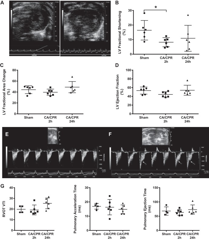
Fig. 1.
Cardiac function 2 h and 24 h after cardiac arrest and cardiopulmonary resuscitation (CA/CPR). A: representative end-diastolic transthoracic short-axis view from sham (left) and post cardiac arrest (right) mouse, 2 h after resuscitation. B: left ventricular (LV) fractional shortening was significantly decreased 2 h after, but not 24 h after CA/CPR. C and D: fractional area change and LV ejection fraction were not significantly different from sham 2 h after or 24 h after CA/CPR. E and F: Doppler trace from the proximal pulmonary artery (PA) 2 h after sham (E) and CA/CPR (F). Arrows point to midsystolic flow reduction (“notching”) in the trace, indicating abnormal right ventricular afterload. G: right ventricular outflow tract (RVOT) velocity-time integral (VTI), pulmonary acceleration time, and pulmonary ejection time were not significantly different between groups.

