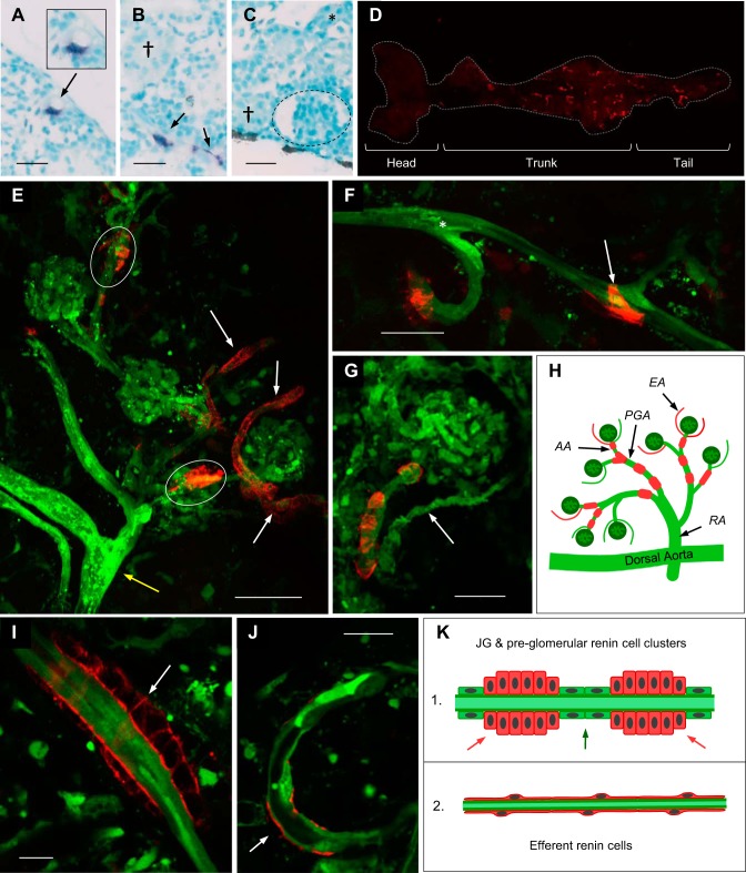
Fig. 1.
Distribution and morphology of mesonephric renin cells. The location and morphology of mesonephric renin cells was assessed across the whole kidney by in situ hybridization and Tg(ren:LifeAct-RFP, kdrl:EGFP). A–C: in situ hybridizations of renal tissue with background structures stained by methyl green and ren mRNA detected by NBT (blue). Perivascular ren is associated with intrarenal vessels (A and B) and not detected in †proximal or *distal tubules or inside glomeruli (dashed outline). Scale bars = 20 µm. GFP and RFP fluorescently label endothelial and renin cells, respectively. D: ventral view of the whole adult kidney in Tg(ren:LifeAct-RFP) showing prominent and sparse expression of ren:LifeAct-RFP in the trunk and tail regions compared with the head kidney, respectively. E–G: maximum-intensity projections of Tg(ren:LifeAct-RFP, kdrl:EGFP). E: group of glomeruli and associated vasculature showing ren:LifeAct-RFP at the afferent arterioles (white ovals) and weaker ren:LifeAct-RFP at the efferent arterioles (white arrows). A larger preglomerular artery is indicated by the yellow arrow. Scale bar = 50 µm. F: expression of ren:LifeAct-RFP at preglomerular arteries. The white arrow shows a branch to an efferent arteriole with ren:LifeAct-RFP, and the asterisk shows a branch without ren:LifeAct-RFP. G: juxtaglomerular (JG) ren:LifeAct-RFP at the afferent arteriole. Scale bars = 25 µm. H: schematic showing localization of renin cells (red) in the renal vasculature (green); RA, renal artery; PGA, preglomerular artery; AA, afferent arteriole; EA, efferent arteriole. I and J: single 1-µm optical sections of Tg(ren:LifeAct-RFP, kdrl:EGFP). I: cross section of multicell epithelioid renin cluster at a preglomerular artery. Boundaries of cuboidal-shaped renin cells are demarcated by ren:LifeAct-RFP. J: cross section of an efferent arteriole showing the thin and small cell body (arrow) of an efferent perivascular renin-expressing cell. Scale bars = 10 µm. K: schematic showing the cross sections of 1) JG and preglomerular renin cell clusters (red arrows) with intermediate smooth muscle cells (green arrow) and 2) efferent arteriolar renin cells. JG and preglomerular renin cells are present as multicellular clusters. Efferent renin cells surround the endothelium with thin-bodied cells that have a low cytoplasmic volume.

