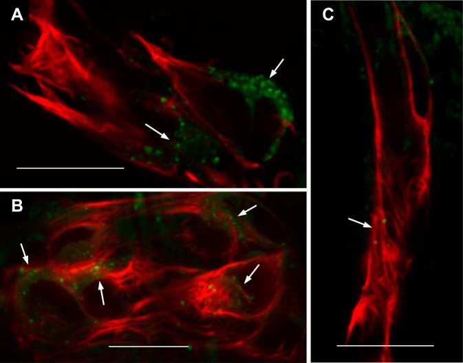
Fig. 3.
Presence of acidic intracellular vesicles in renin cells. To test for the presence of acidic granules in renin cells across the mesonephric kidney, whole kidney squashes of Tg(ren:LifeAct-RFP) were stained with LysoTracker Green. Single 0.5-µm optical sections were taken by confocal microscopy to assess staining in ren:LifeAct-RFP-expressing cells. A: a juxtaglomerular ren-RFP cell cluster with regions of punctate intracellular LysoTracker staining (white arrows). B: preglomerular arteriolar renin cell cluster also with regions of punctate LysoTracker staining (white arrows). C: efferent arteriole showing very occasional LysoTracker-stained vesicles (arrows). All scale bars = 10 µm.

