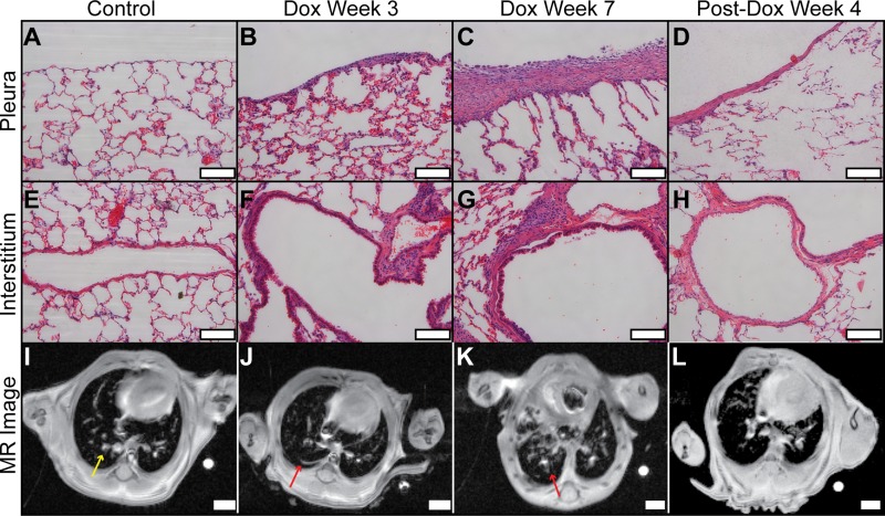
Fig. 2.
Comparison of histology and MRI. Histology scale bars (A−H) are 100 μm (×20 magnification); MR image scale bars (E−H) are 1 mm. The pleura of the control (A) was thin but was progressively thicker in animals treated with Dox for 3 and 7 wk (B and C, respectively). Four weeks after Dox treatment (D), pleural thickening partially resolved. Similar histological patterns are observed for vascular-adventitial fibrosis (I−L). In the MR images from the same animals, the control (I) displayed low signal from lung parenchyma and high signal from larger pulmonary vessels (e.g., yellow arrow). In contrast, animals undergoing Dox treatment (J and K) increasingly displayed high signal structures that extended from the subpleural region and adventitia into the interstitium (e.g., red arrows). High signal volume decreased 4 wk after Dox treatment ended (L), consistent with progression and resolution patterns observed with conventional histology.

