Fig. 2.
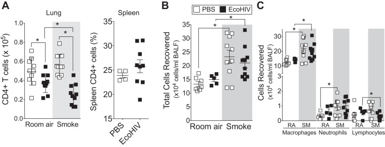
Animals infected with EcoHIV have altered lung immune cell profile following cigarette smoke exposure. A/J mice were inoculated one time with EcoHIV or PBS. Later (1 mo), the animals began daily exposures to cigarette smoke. Mice were euthanized at 2 mo. A: absolute lung CD4+ cell numbers were quantified by flow cytometery. The percentage of spleen CD4+ T cells was also quantified in animals infected with EcoHIV for 3 mo. B and C: total bronchoalveolar lavage fluid (BALF) cells (B) and macrophages, neutrophils, and lymphocytes (C) were quantified in each animal group by Quik Diff analysis of BALF cells following cytospin. Graphs are represented as means ± SE of 3 separate measurements on independent days. *P <0.05 when comparing both treatments connected by a line, determined by 2-way ANOVA with Tukey's post hoc test; n = 10 experiments/group.

