
Fig. 4.
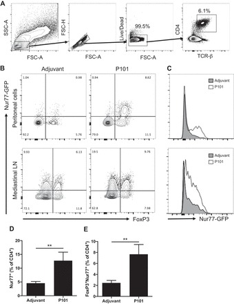
Activation of regulatory T cells (Tregs) and other CD4+ T cells in mice vaccinated with P101 peptide. A: peritoneal cavity and mediastinal lymph nodes were harvested from Nur77GFP mice immunized with P101 or adjuvant only and analyzed by multicolor flow cytometry at 24 hrs. The analyzed populations were gated on live singlet TCRβ+CD4+ T cells. B: representative plots of Nur77 (by GFP) and FoxP3 (by PerCP-Cy5.5 labeled antibody) expression on peritoneal CD4+ T cells (top) and mediastinal lymph node CD4+ T cells (bottom). C: histogram of Nur77-GFP expression among FoxP3+CD4+ T cell from peritoneal cavity (top) and mediastinal lymph node (bottom). Each plot is representative of 4 independent experiments. Percentage of Nur77+ cells (D) and FoxP3+Nur77+ cells (E) among peritoneal CD4+ T cells. n = 5 in each group. **P < 0.01. Data are means ± SE. Significance was assessed by Mann-Whitney test.