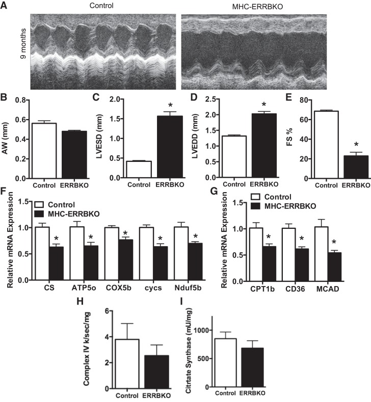
Fig. 3.
Noninvasive ECHOs of MHC-ERRBKO. A: sample M-mode echocardiograms from the left ventricle (LV). B: anterior wall thickness (AW). C: left ventricular end systolic diameter (LVESD). D: left ventricular end diastolic diameter (LVEDD). E: percent fractional shortening (%FS). F: qPCR expression of OXPHOS genes. G: qPCR expression of FAO genes. H: complex IV enzymatic activity. I: citrate synthase activity of MHC-ERRBKO and control animals. Data are presented as means ± SE; n = 4–6 per group; *P < 0.05 compared with control animals.

