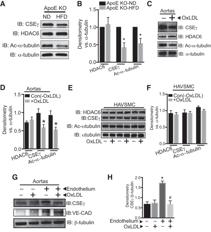
Fig. 4.
A and B: isolated aortas from ApoE KO mice fed HFD or ND for 12 wk were subjected to Western blotting using CSEγ, acetylated α-tubulin, total α-tubulin, HDAC6, and β-tubulin antibodies. C and D: isolated aortic segments were incubated in medium with or without OxLDL (50 µg/ml) for 24 h, and cell lysates were subjected to Western blotting using CSEγ, acetylated α-tubulin, total α-tubulin, and HDAC6 antibodies. E and F: human aortic vascular smooth muscle cells (HAVSMC) were incubated in medium with or without OxLDL (50 µg/ml) for 24 h, and cell lysates were subjected to Western blotting using CSEγ, acetylated α-tubulin, total α-tubulin, and HDAC6 antibodies. G and H: CSEγ and VE-cadherin (CAD) protein expression were determined by Western blotting in control and deendothelialized mouse aortas in the presence or absence of OxLDL (50 µg/ml) for 24 h. *P < 0.05 vs. respective controls. #P < 0.05 vs. endothelium-intact group not treated with OxLDL. Western blots are representative of 3 immunoblots.

