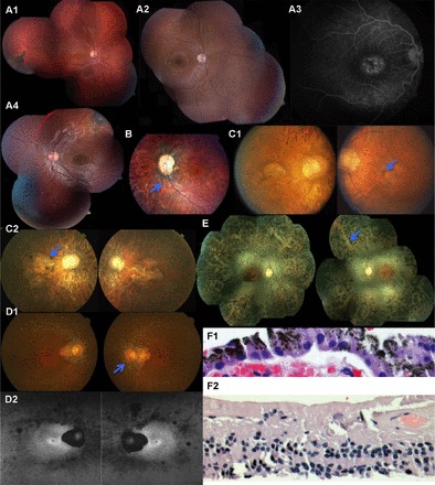
Fig. 4.

A1: fundus examination of RF.H.0909: III-I at age 11 yr. A2 and A3: the fundus image and fluorescein angiography of RF.H.0909: III-II. A4: the fundus image of patient RF.H.0909: III-III. B: fundus photos of patient RF.T.1111: II-V showing optic disk pallor with an optic pit temporally retinal vascular attenuation and bone spicule pigmentary change. C1 and C2: the color fundus photos of the patient RF.S.0711: II-I at age 36 and 50 yr. D1: color fundus photos at age 59 yr; D2: autofluorescence (AF) fundus photos of patient RF.SH.0008: II-III at age 63 yr demonstrate mottled areas of reduced AF most prominent along arcades and nasal midperiphery, a large patch of complete lack of AF associated with peripapillary chorioretinal atrophy, and small perifoveal hyper-AF ring oculus uterque (OU). E: fundus examination of RF.M.0711: II-I showed mild optic nerve pallor, attenuation of retinal blood vessels. F1 and F2: hematoxylin-and-eosin stain of retinal biopsy of RF.PO.1109: II-I reveals retinal degeneration of all layers including retinal ganglion cell, inner nuclear layer, and outer nuclear layer and intact retinal pigment epithelial (RPE) monolayer above choriocapillaris.
