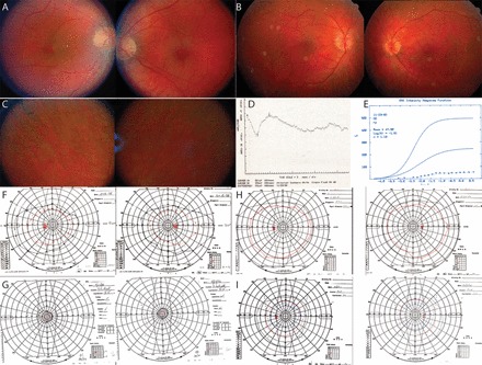
Fig. 5.

Pedigree RF.TE.0512 with DHDDS c.124A>G; p.Lys42Glu mutations. Top: color fundus photos at age 22 yr for II-I (A) and II-II (B) show mild disk pallor and retinal vascular attenuation. Middle: color fundus photos show peripheral bone spicule pigment and white spots in II-I at age 22 yr (C). Full-field electroretinogram (ERG) shows reduction of a-wave and b-wave responses to a scotopic bright flash (D); an intensity-response curve shows reduced maximum amplitude (Rmax) (E). Bottom: Goldmann visual fields at age 22 yr (F) and age 40 yr (G) in II-I and at age 22 yr (H) and age 32 yr (I) in II-II show progressive constriction.
