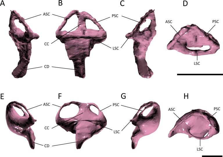
Figure 6. Endosseous labyrinth.
(A–D) left inner ear of Pelagosaurus typus and (E–H) left inner ear of Gavialis gangeticus. (A–E) anterior view; (B–F) lateral view; (C–G) posterior view; (D–H) dorsal view. Abbreviations: ASC, anterior semicircular canal; CC, common crus; CD, cochlear duct; LSC, lateral semicircular canal; PSC, posterior semicircular canal. For visualization, the labyrinth of Gavialis has been scaled to the same anteroposterior width as Pelagosaurus. Scale bars equal 1 cm.

