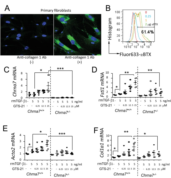
Figure 11.
Effects of α7 nAChR activation on TGF-β1–induced lung profibrotic genes in mouse lung primary fibroblasts. (A) Immunofluorescence of mouse lung primary fibroblasts. The cultured mouse lung primary fibroblasts were labeled with or without rabbit anti-collagen 1 antibody. The stained cells were subjected to confocal microscopy. Green: FITC, collagen 1 positive; blue: DAPI, nucleus. (B) Flow cytometry analysis of α7 nAChR expression in mouse lung primary fibroblasts. The cells were collected and stained with fluor-633 α-BTX at 0, 0.5, 1 and 2 μg/mL. (C–F) Activation of α7 nAChR affects profibrotic genes in rmTGF-β1–stimulated fibroblasts 24 h after rmTGF-β1 challenge. The cells were pretreated with GTS-21 (6.25, 12.5 and 25 μM separately; PBS was applied in control group) 30 min before stimulation with rmTGF-β1 (5 ng/mL). The cells were harvested 24 h after rhTGF-β stimulation. (C) Chrna7 mRNA. (D) Fstl1 mRNA. (E) Acta2 mRNA. (F) Col1a1 mRNA. *p < 0.05, **p < 0.01, ***p < 0.001 in compared groups. N = 3–6 in each group. Data are mean ± SD. Data were pooled from two independent experiments.

