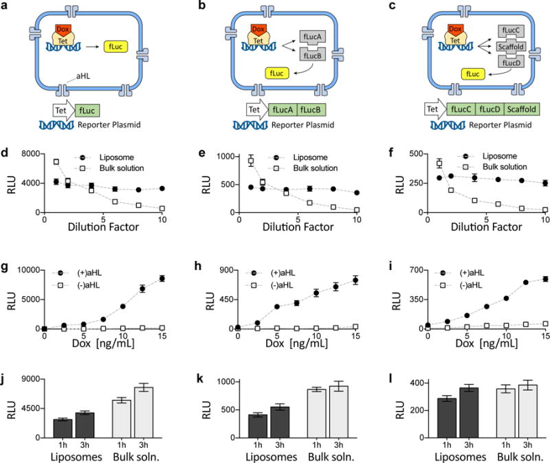
Fig 3. Comparison of single- and multi-component genetic circuits.

a–c. Genetic cascades involving one-, two-, or three-part luciferase protein assemblies. Expressed under doxycycline-inducible Tet promoters were whole firefly luciferase (fLuc) (a), the two halves (here denoted fLucA and fLucB) of split fLuc bearing split inteins and mutually binding coiled coils (b), and two halves (here denoted fLucC and fLucD) of split fLuc bearing split inteins and coiled coils that bind to a third common template (denoted “scaffold”) (c). d–f. Effects of dilution on fLuc expression in liposomes vs. bulk solution, for the fLuc assemblies described in a–c (see Fig. S6 for experiments under the control of a constitutive P70 promoter). Dotted lines throughout this figure are visual guides, not fits. RLU, relative light units. g–i. End-point expression of luciferase measured at the 3 h time point, for 7 different concentrations of doxycycline (Dox). See Fig. S7 for corresponding 1 h end-point expression data, and Figs. S8 – S10 for the same reactions in bulk solution. j–l. Comparison of liposomal vs. bulk solution expression of luciferase, at 2 different time points and for 10 ng/mL of Dox. The 2 plasmids in k and 3 plasmids in l were mixed at equimolar ratios, with total DNA concentration held constant. Error bars indicate S. E. M. n=4 replicates.
