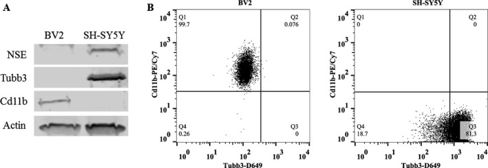
Figure 1.

BV2 express microglial markers and SH‐SY5Y express neuronal markers, indicating they are microglial‐like and neuronal‐like cells, respectively. (A) Western blot analysis of basal expression of microglial and neuronal cell type markers in cell lysates of BV2 and SH‐SY5Y. SH‐SY5Y were differentiated into a neuron‐like phenotype using retinoic acid (RA) for 4 days. Microglia‐like BV2 express microglial marker Cd11b. Neuron‐like SH‐SY5Y express neuronal markers neuron‐specific enolase (NSE) and β‐III tubulin (Tubb3). β‐actin was used as a loading control. (B) Flow cytometry analysis of basal expression of microglial and neuronal markers in BV2 and SH‐SY5Y. The majority of BV2 cells express microglial marker Cd11b, whereas the majority of SH‐ SY5Y express neuronal marker Tubb3. n = 3 per group. Gates were established using unstained and isotype controls for each antibody.
