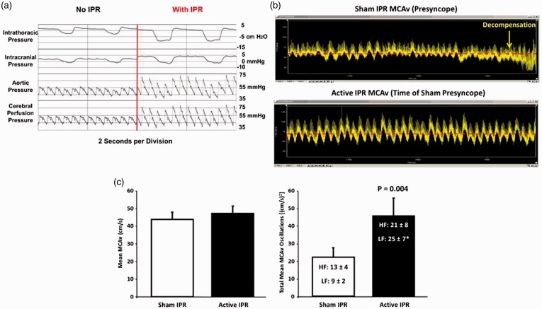
Figure 8.
(a) Results from experiments conducted using an animal (porcine) model of 40% controlled hemorrhage demonstrate that reduced intrathoracic pressure lowers right atrial pressure, which in turn pulls more venous blood back into the thorax and increases arterial pressure and reduces intracranial pressure, which together increase cerebral perfusion. Modified from Convertino et al.17 (b) Arterial waveform recordings demonstrate that a delay in the onset of symptoms and hemodynamic decompensation is associated with increases in CBFV oscillations when intrathoracic pressure is reduced by breathing against resistance (bottom recording) compared to breathing with normal intrathoracic pressure in the same subject (top recording). Modified from Convertino et al.16 (c) Individuals with high tolerance to central hypovolemia (closed bars) display greater oscillations in mean medial cerebral artery blood flow velocity (MCAv) at the onset of decompensation (right panel) compared to individuals with low tolerance (open bars) despite similar levels of mean blood flow (left panel). Symbols are mean ± 1 sem. Modified from Rickards et al.32 (A color version of this figure is available in the online journal.)

