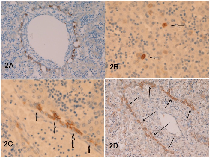
Figure 2.
(a) NCAM is always expressed in some cells of ductal plate (9 gestational weeks (GW)). (b) KIT expression is seen in some hepatoblasts and hematopoietic progenitor cells of the parenchyma (arrows, 12 GW). (c) The cells of ductal plate (arrows) are strongly positive for MET (9 GW). (d): The cells of remodeling ductal plate (arrows) indicated by arrows are strongly positive for PDGFRA (13 GW). The staining pattern is mostly membranous. (a, d) × 200. (b, c) × 400. (A color version of this figure is available in the online journal.)

