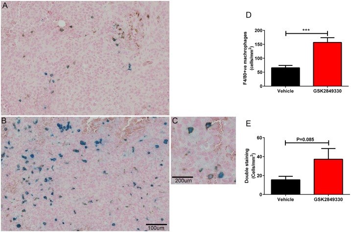
Fig 7. Immunohistochemistry analysis of tumors from the USPIO MRI study.
Panels A and B show Prussian blue and F4/80 IHC staining for vehicle and GSK2849330 groups respectively. A region of tumor reflecting cytoplasmic iron staining coregistered with a sub population of plasma membrane F4/80 positive (F4/80+ve) cells is observed in C. The quantitative data (cells/mm2) reflects an increase in number of F4/80+ve macrophages (D) and a trend toward increased coregistered F4/80+ve macrophages with USPIO (E) in the GSK2849330 treated group compared to the vehicle group. Data is presented as mean±SEM (S5 Table). ***P<0.01: unpaired t-test (two-tailed).

