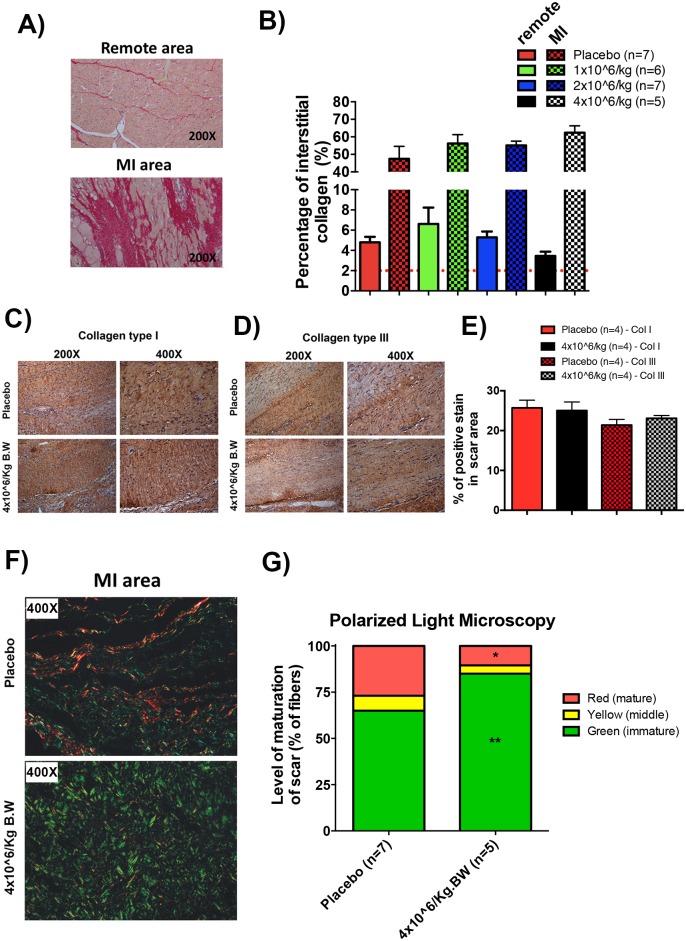
Fig 4. Injection of 4 million cell/Kg increased the deposition of immature fibers in the scar post-MI.
A) Representative images of septum (remote area) and posterior wall (MI-border transition area) of left ventricle stained with PicroSirius-red. B) quantification of interstitial collagen in the septum (remote area) and posterior wall (MI-border transition area). C) representative immunohistochemistry images of MI scar area, showing staining of collagen fibers types I D) and type III, in placebo and 4 million cell/Kg injection groups and E) quantification of the percentage of positive stain of collagen types for both group. F) representative polarized light microscopy (non-antibody reaction) images of scar areas showing collagen mature fibers in red and immature fibers in green for both placebo and 4 million cell/Kg injection groups and G) quantification of collagen fiber maturity in scar area for both placebo and 4 million cell/Kg injection groups (differences are significant between 4 million cell/Kg injection and placebo group * p<0.05 to mature (red) fibers and ** p<0.005 to immature (green) fibers, Student’s t-test to each maturity fibers). Data are mean ± SEM.

