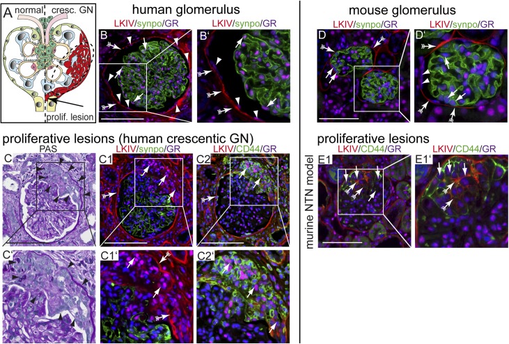
Figure 1.
GR expression in proliferative lesions. (A) Schematic of a glomerulus with a proliferative lesion (i.e., cellular crescent in crescentic GN; right). Obstruction of the tubular outlet by a crescent (arrow) triggers irreversible loss of the nephron and renal function. Blue, podocytes; light green, quiescent PECs. (B and B′) Immunofluorescence staining of a normal human glomerulus (LKIV stains PEC matrix of Bowman’s capsule in red; arrows with tails), podocytes (antisynaptopodin; green), and the GR (magenta). GR expression is visible in all glomerular cells, including podocyte (arrow) and PEC nuclei (arrowheads). (C–C2) Serial sections of a human biopsy with an extracapillary proliferative lesion (arrowheads in C and C′; Goodpasture syndrome). (C1) GR was expressed at low levels in podocytes, which were rarely observed in cellular crescents. GR colocalized with PEC activation marker CD44 (arrows in C2) and PEC matrix (LKIV; arrows with tails). GR (magenta) is expressed in all cell nuclei within sclerotic lesions. GR expression in murine glomerulus. Scale bars, 100 μm. (D and D′) Normal mouse glomerulus (red; PEC matrix [LKIV]; arrows with tails), podocytes (antisynaptopodin; green), and GR (magenta) show ubiquitous nuclear staining of the GR in all glomerular cells. Activated PECs express the GR in experimental GN in mice in proliferative and sclerotic lesions. (E1) Crescentic nephritis model (NTN): CD44–positive activated PECs (green; arrows) deposit PEC matrix (LKIV; red; arrows with tails) and populate a cellular lesion. Scale bars, 50 μm.

