Figure 3.
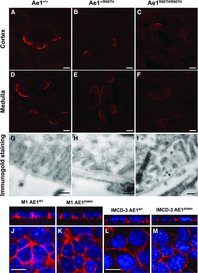
The R607H/R589H variant is correctly targeted to the basolateral plasma membrane. (A–F) High magnification images of cortical CD (A–C) and inner stripe of the outer medulla (D–F) in Ae1+/+, Ae1+/R607H, and Ae1R607H/R607H showing that Ae1 R607H is localized to the basolateral pole of A-ICs. Scale bar: 10 µm. (G–I) The basolateral targeting of Ae1 R607H is evident from immunogold labeling in cortical A-ICs. Scale bar: 250 nm. (J–M) M1 (J and K) or mIMCD-3 (L and M) cells expressing either human kAE1 WT-myc (J and L) or R589H-HA (K and M) were grown to confluency for 7 days on semipermeable filters before fixation, permeabilization, and detection of kAE1 with either mouse anti-myc or anti-HA antibody, followed by Cy3-coupled anti-Ig (red). Nuclei were stained with DAPI. Scale bar: 10 µm.

