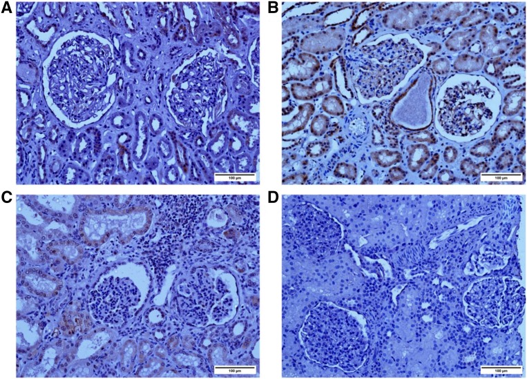
Figure 2.
MAGI2 expression is altered in patients with mutations. Immunohistochemical staining with human anti–MAGI2 antibody (Sigma-Aldrich) is shown. (A) A kidney that was not suitable for transplant was used as a control. (B) Biopsy specimen obtained from an individual with minimal change disease; MAGI2 staining is seen in the glomeruli at the periphery of capillary loops, consistent with podocyte localization. Staining in tubules is also seen. (C) Nephrectomy specimen obtained from patient 175 (homozygous mutation in MAGI2) shows weak but positive glomerular MAGI2 staining. (D) Biopsy specimen obtained from patient 180 (compound heterozygous mutation in MAGI2) shows complete lack of MAGI2. Original magnification, ×20. Scale bar, 100 μm.

