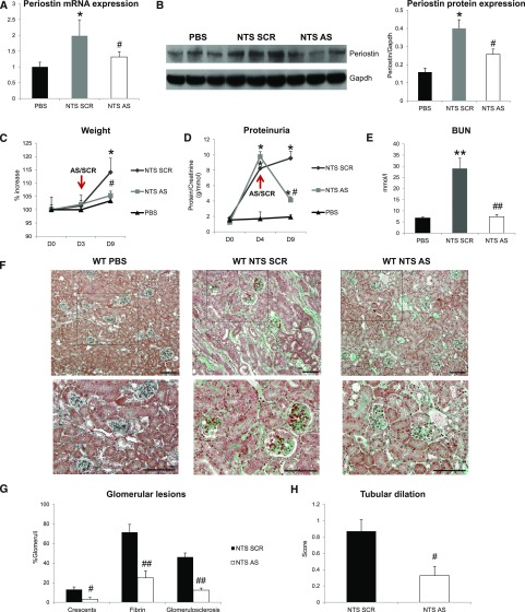
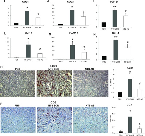
Figure 3.
Delayed treatment with antisense against periostin reverses the development of NTS-induced GN. (A and B) Periostin mRNA (A) and protein (B) is increased in NTS scrambled mice but is decreased in mice treated with antisense ODN. (C–E) Initiation of antisense treatment at D3 after NTS reversed the increase in weight (C), proteinuria (D), and BUN (E) observed in NTS scrambled mice. (F) Masson trichrome staining showing reduced fibrosis and tissue damage in NTS antisense-treated mice (upper panels, ×200; lower panels, ×400). (G and H) Quantification of structural glomerular (G) and tubular (H) alterations showing highly reduced crescent formation, fibrin deposition, glomerulosclerosis, and tubular dilation in NTS antisense-treated mice. (I–K) mRNA expression of the fibrotic molecules COL1 (I), COL3 (J), and TGF-β1 (K) is attenuated in antisense-treated mice after NTS. (L–N) mRNA expression of the inflammatory mediators MCP-1 (L), VCAM-1 (M), and CSF-1 (N) is diminished in antisense-treated mice after NTS. (O and P) F4/80 (O) and CD3 (P) staining showing markedly decreased accumulation of macrophages and lymphocytes, respectively, in NTS mice treated with antisense against periostin. Quantification of the staining is shown in the graphs on the right. Animals were euthanized after 9 days of NTS administration. Scale bars, 100 μm (F, O, and P). n=5 per group. *P<0.05; **P<0.01 versus PBS; #P<0.05; ##P<0.01 versus NTS scrambled.


