Figure 6.
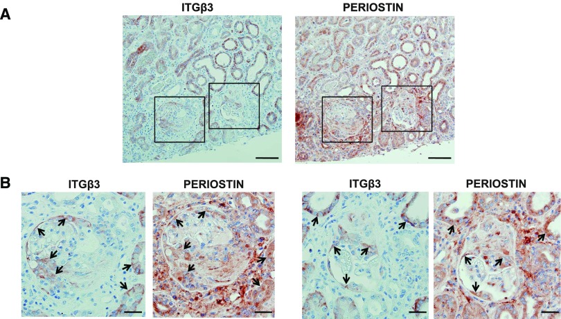
Periostin and integrin-β3 are coexpressed in human renal biopsies. (A) Immunohistochemical staining of integrin-β3 and periostin in consecutive sections from a biopsy specimen of a patient with ANCA vasculitis, depicting a strong expression and a high degree of colocalization between periostin and integrin-β3 both in damaged glomeruli and tubules. (B) Magnified images of the panels drawn in (A), showing in more detail the coexpression of periostin and integrin-β3 in damaged glomeruli and surrounding tubules. Arrows indicate colocalization sites. Scale bars, 100 μm (A) or 30 μm (B). Representative images are shown.

