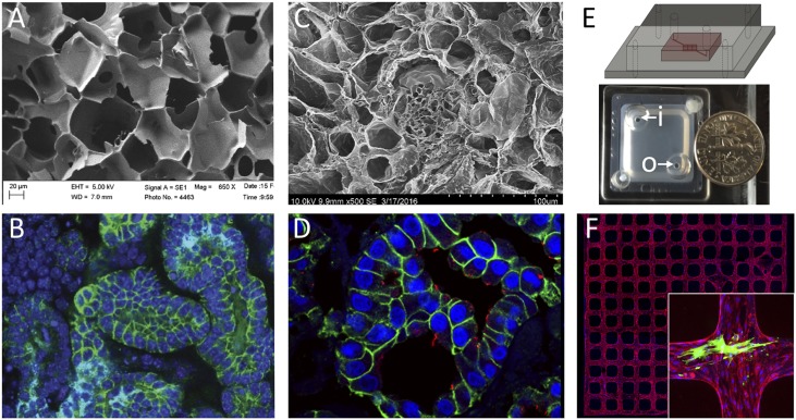
Figure 3.
Multiple different bioengineering approaches to generating tissue scaffolds offer unique advantages for rebuilding kidney tissue. Polymer-based (A and B), decellularized tissue (C and D), and organ-on-a-chip (E and F) approaches are shown. (A) Scanning electron micrograph of 6% silk scaffold before cell seeding; a network of channels is formed into which NPCs can be seeded. Photo: Jeannine Coburn. (B) E-cadherin (epithelial cells) and DAPI (nuclei) staining reveals a network of tubules with lumens within the silk approximately 2 weeks after seeding NPCs. Photo: Jessica Davis-Knowlton. (C) Scanning electron micrograph of decellularized kidney tissue showing the extracellular matrix framework for tubules and glomeruli. Photo: Joseph Uzarski. (D) Immunostaining of a decellularized kidney that has been recellularized with MDCK cells 7 days after cell seeding. E-cadherin (green) and luminal cilia (acetylated tubulin, red) demonstrate epithelialization. Photo: Joseph Uzarski. (E) Schematic and top view picture of a vascular network-on-a-chip device that allows flow through the network via inlet (i) and outlet (o). Photo: Zheng laboratory. (F) An example view of a three dimensional microvessel network formed by mouse kidney endothelial cells. Red: CD31, blue: DAPI. The inset shows fluorescence immunostaining of a device in which podocytes (green) were cocultured with the vascular endothelial network (red). Photo: Zheng laboratory. EHT, extra high tension voltage setting; WD, working distance.

