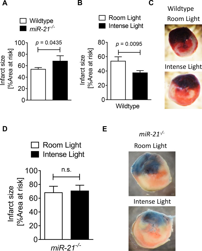
Fig 5. Light elicited cardioprotection in wildtype and miR-21-/- mice.
(A-E) Mice underwent 60 min of ischemia and 120 min of reperfusion at room light (200LUX) or after exposure to 3 hours of intense light (10,000 LUX). Infarct sizes were measured by double staining with Evan’s blue and triphenyl-tetrazolium chloride. Infarct sizes are expressed as the percent of the area at risk (AAR) that underwent infarction. (A) Infarct sizes in wildtype or miR-21-/- mice at room light conditions (mean±SD, n = 4, p<0.05). (B, C) Infarct sizes in wildtype mice after exposure to intense light for 3 h compared to room light conditions. (mean±SD, n = 4, p<0.05). (C) Representative infarct staining in hearts from wildtype mice exposed to intense light or room light prior to in situ myocardial ischemia and reperfusion (blue, retrograde Evan’s blue staining; red and white, area at risk; white, infarcted tissue). (D, E) Infarct sizes in miR-21-/- mice exposed to intense light or room light prior to in situ myocardial ischemia followed by reperfusion (mean±SD, n = 4, not significant). (E) Representative infarct staining in hearts from miR-21-/- mice exposed to intense light or room light prior to in situ myocardial ischemia reperfusion (blue, retrograde Evan’s blue staining; red and white, area at risk; white, infarcted tissue).

