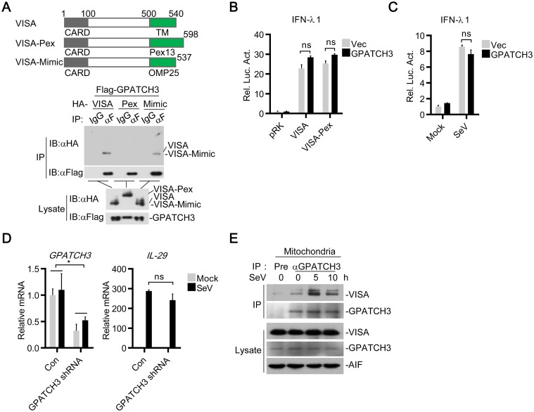
Fig 6. GPATCH3 targets mitochondria localized VISA.
(A) 293T cells (2 x 10⁶) were cotransfected with Flag-GPATCH3 (3 μg) and HA-tagged VISA, VISA-Pex, or VISA-Mimic (3 μg). Coimmunoprecipitation and immunoblotting were performed with the indicated antibodies. (B) 293T cells (1 x 10⁵) were cotransfected with IFNλ1 reporter (0.05 μg), the indicated expression plasmids (0.05 μg each) and empty vectors or GPATCH3 expression plasmids (0.05 μg). Luciferase assays were performed 24 hours after transfection. (C) 293T cells (1 x 10⁵) were transfected with the IFNλ1 reporter (0.05 μg) together with empty vectors or GPATCH3 expression plasmids (0.05 μg). Twenty-four hours after transfection, cells were stimulated with SeV for 24 hours before luciferase assays were performed. (D) 293T cells (4 x 10⁵) were transfected with control- or GPATCH3-shRNA plasmids (1 μg). Thirty-six hours later, cells were left uninfected or infected with SeV for 6 hours before total RNAs were extracted and the mRNA levels of the indicated genes were analyzed by qPCR. (E) 293T cells (1.5 x 10⁷) were left uninfected or infected with SeV for the indicated times. Mitochondria were isolated by subcellular fractionation and the mitochondrial lysates were subjected to co-immunoprecipitation and immunoblotting analysis with the indicated antibodies. Graphs show mean ± SD. n = 3. *P<0.05, **P<0.01 (Student’s t-test).

