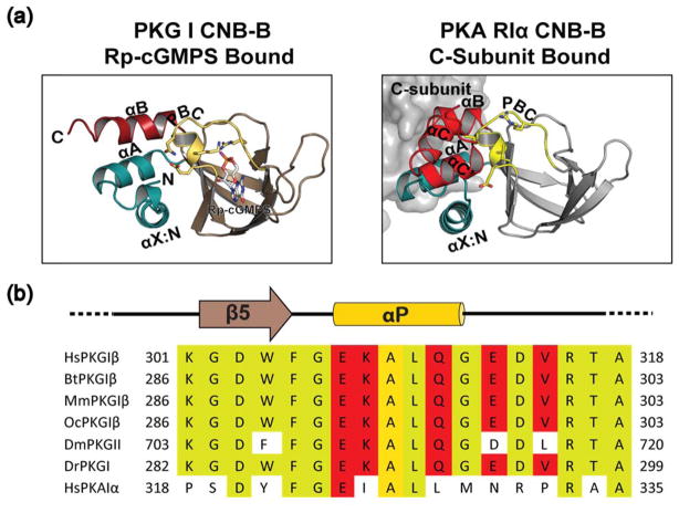
Fig. 4.
Comparison of PKA RIα:C complex with PKG Iβ CNB-B Rp-cGMPS structure. (a) structures of PKG Iβ CNB-B:Rp-cGMPS, left and the CNB-B of PKA RIα:C holoenzyme (PDB ID:2QCS), right. Same color scheme as Fig. 1, C-subunit shown as a grey surface. (b) sequence alignments of PKG Iβ in various species and human PKA RIα. Yellow shading indicates conserved residues and red shading those residues which stabilizes the apo like state when bound to Rp-cGMPS.

