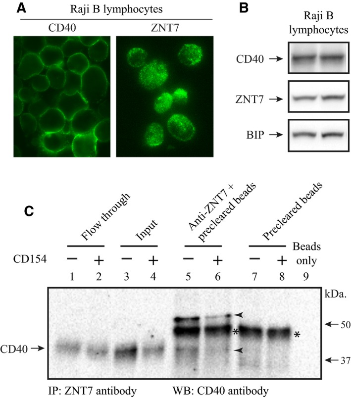
Figure 3.

Interaction of ZNT7 with CD40 in Raji B lymphocytes. (A) Subcellular localization of endogenous ZNT7 and CD40 in Raji B cells. Cells were seeded in the complete RPMI 1640 medium without FBS for 2 h before staining. ZNT7 and CD40 were detected by antibodies against ZnT7 23 and CD40, respectively. (B) Expression of endogenous ZNT7 and CD40. Proteins were detected using anti‐ZnT7 and anti‐CD40 antibodies by western Blot analysis. The expression of BIP was used as the loading control. The two lanes are the protein lysate harvested from two independent experiments. (C) Immunoprecipitation. Raji B cells treated with or without CD154 (100 ng·mL−1) were harvested for the immunoprecipitation assay as described in Materials and methods. CD40 was immunoprecipitated by the antibody against human ZNT7 and detected by the antibody against CD40 in western blot analysis. Interaction of ZNT7 with CD40 was observed in lanes 5 and 6 (arrowheads). Two CD40 protein bands were pulled down by the ZNT7 antibody, ~ 40 and ~ 60 kDa. The lower band is the intact CD40 protein and the upper band may represent an additional protein bound to the ZNT7‐CD40 complex. The asterisk represents a nonspecific binding of protein to the goat anti‐(rabbit IgG) magnetic beads. IP, immunoprecipitation; WB, western blot assay. Molecular markers are indicated by arrows. The amounts of the input (lanes 3 & 4) were 1.6% of the starting lysate used in the IP assay.
