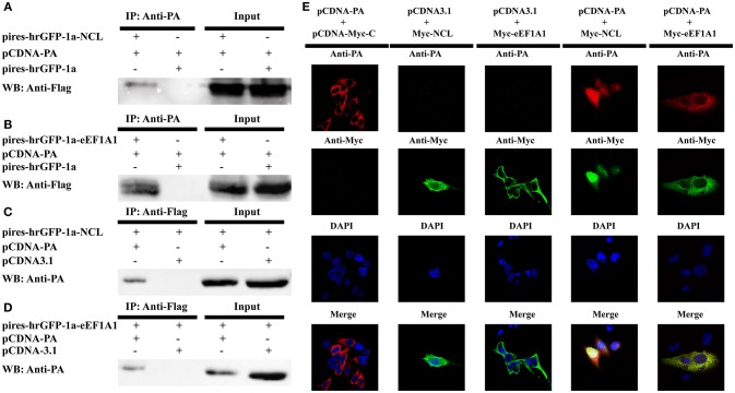
Figure 7.
Confirmation of the interaction of IAV PA with NCL and eEF1A1. 293T cells were transiently co-transfected with the PA-expressing plasmid or the empty vector and the tagged NCL, eEF1A1 or the empty vector for co-immunoprecipitation and co-localization studies. (A–D) Cell lysates were prepared at 48 h post-transfection and the proteins were immunoprecipitated with anti-PA or anti-FLAG antibodies. Proteins in cell lysates (input) and immunoprecipitated samples were detected with the antibodies against FLAG or PA in Western blot. (A) Co-precipitation of about 110 kDa NCL protein with recombinant viral PA in cell lysate. (B) Co-precipitation of about 50 kDa eEF1A1 protein with PA in cell lysate. (C) Co-precipitation (reverse IP) of about 85 kDa PA with NCL. (D) Co-precipitation (reverse IP) of about 85 kDa PA with eEF1A1. (E) Confocal microscopy analysis was carried out for demonstrating colocalization of PA and NCL or eEF1A1. 293T cells were transiently co-transfected with PA expressing vector or empty vector and Myc-tagged NCL expressing vector, eEF1A1 expressing vector or empty vector, respectively. 24 h later, cells were fixed, and yellow regions are the areas of PA and NCL or eEF1A1 co-localization. Nuclei were stained using DAPI (blue).

