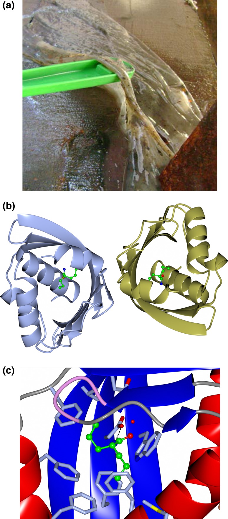
Fig. 2.
a The microbiological mat at the Tomsk sampling site, Parabel, Tomsk Region, West Siberia, Russia, which provided the metagenome DNA sample where the LEH was identified. Picture kindly provided by Prof. Elizaveta Bonch-Osmolovskaya. b A cartoon representation of the dimeric Tomsk-LEH structure in complex with an inhibitor valpromide shown in ball and stick representation which is bound at the active site. PDB code 5IG. c A close-up representation of the active site of Tomsk-LEH with the active site residues and inhibitor highlighted. The red sphere represents the active site water molecule. Images were generated using CCP4 MG [48]

