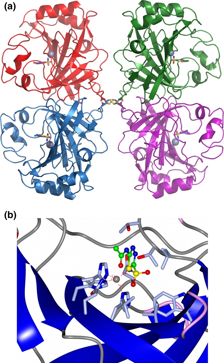
Fig. 3.
a A cartoon representation of the tetramer structure of the α-carbonic anhydrase from T. ammonificans showing each subunit in a different colour. The active site zinc is shown as a sphere in each subunit together with an inhibitor in ball and stick representation bound at each active site. The disulfide bonds at the centre of the tetramer which add stability to the enzyme are shown in yellow. b A close-up representation of the active site of the enzyme showing the catalytic zinc molecule and the inhibitor acetazolamide bound to the active site. Side chain residues co-ordinating the zinc ion and residues making up the active site are highlighted. PDB code 4COQ. Images were generated using CCP4 MG [48] and PyMOL [15]

