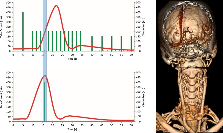
Fig. 1.
Schematic overview of the One-Step Stroke Protocol with 3D volume rendering of the head CTP and volume neck CTA. The One-Step Stroke Protocol consists of a 16-cm volumetric whole-brain CTP acquisition that is interrupted as soon as contrast material is detected in the arteries of the central slab of the 3D volume. Within 1.8 s, the table is then rapidly moved to the neck, where a 16-cm volumetric scan is performed with 0.5 s acquisition time. The table is then automatically moved back to the brain to resume the CTP acquisition. Note that the arterial enhancement in the neck is excellent because the contrast reaches the neck earlier than the brain and the enhancement curve in the neck is shifted to the left

