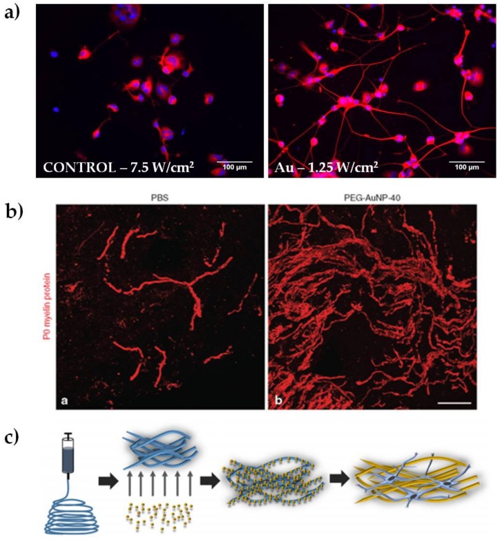
Figure 2.
Representative results of Au NPs for peripheral nerve regeneration. (a) Examples of epifluorescence images of NG108-15 neuronal cells cultured alone or with Au NRs and exposed to different laser irradiances, as indicated in each panel. Cells were marked for β-III tubulin (in red) and DAPI (in blue, reproduced with permission from [49]); (b) Spontaneous remyelination by Schwann cells (myelin marker P0, in red) was enhanced in mice treated with polyethylene glycol-coated Au NPs (reproduced with permission from [7]); (c) schematic representation of electrospun nanofibers doped with 10 nm Au NPs (reproduced with permission from [27]).

