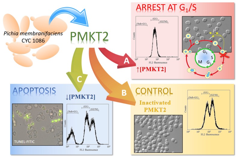
Figure 3.
Mechanisms of the killing activity of PMKT2 depending on toxin dosage. (A) High concentrations of PMKT2 caused cell cycle arrest at G1/S. Flow cytometry analyses of DNA content of S. cerevisiae revealed that cells treated with high doses of PMKT2 were arrested at an early S phase with a nascent bud. (B) Asynchronously growing cells exposed to inactivated toxin remained fully viable. (C) Cells treated with low concentrations of PMKT2 lead to the typical markers of apoptosis. Figure adapted and reproduced with permission (Santos et al. 2013) [42].

