
Figure 1.
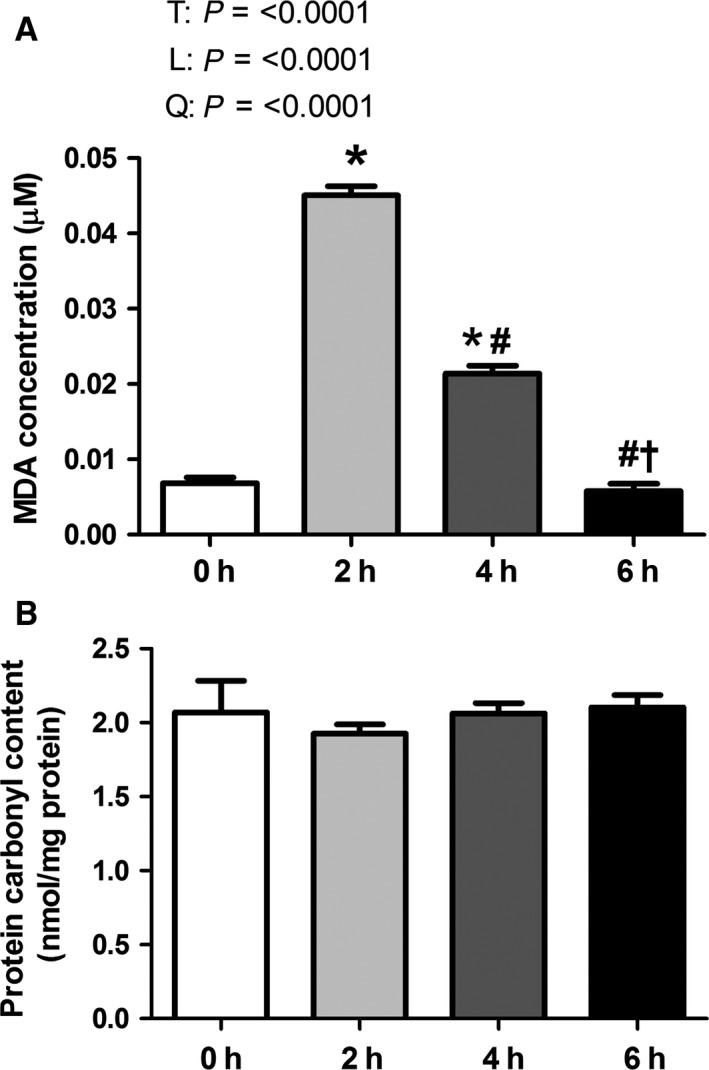
Short‐term environmental hyperthermia increased oxidative stress markers in oxidative skeletal muscle. Following 0, 2, 4, and 6 h of environmental hyperthermia, malondialdehyde (MDA) (A) and protein carbonyl content (B) were measured (n = 8/group). MDA concentration is expressed in μM. Protein carbonyl content is expressed in nmol/mg. * indicates significant difference compared to thermal neutral control, P < 0.05; # indicates significant difference compared to 2 h of heat stress, P < 0.05; † indicates significant difference compared to 4 h of heat stress; T indicates time effect between the treatment, P < 0.05; L indicates linear effect between the treatment, P < 0.05; Q indicates quadratic effect between the treatment, P < 0.05.